Figure 5.
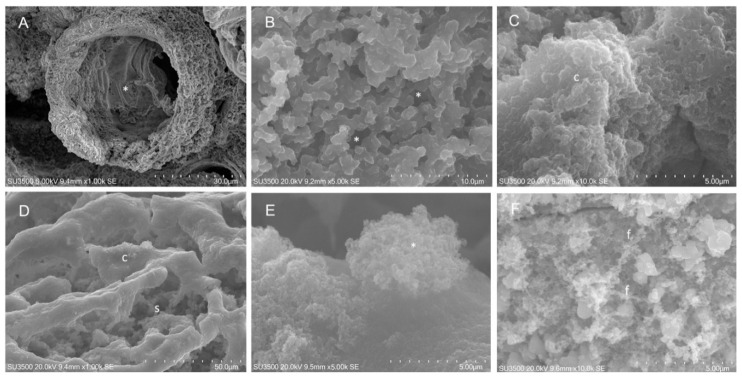
SEM analysis on S. aureus 5S. Untreated S. aureus 5S: (A) SEM, 1000×. Biofilm showed microchannels (asterisk). Biofilm surface was compact and rough; (B) SEM, 5000×. Inner areas with spongy structure. (C) SEM, 10,000×. Sometimes, inner dense areas with compact arrangement were observed. S. aureus 5S after exposure to EO45 1.00% v/v: (D) SEM, 1000×. Compact areas flakes off, allowing inner spongy structure to appear. (E) SEM, 5000×. High magnification showed signs of EPS disintegration in the way of a bush-like floccular aggregate (asterisk). (F) SEM, 10,000×. Very high magnification shows trabeculae flaking in fine filaments. f: filaments; s: sponge; c: caves.

