Figure 4.
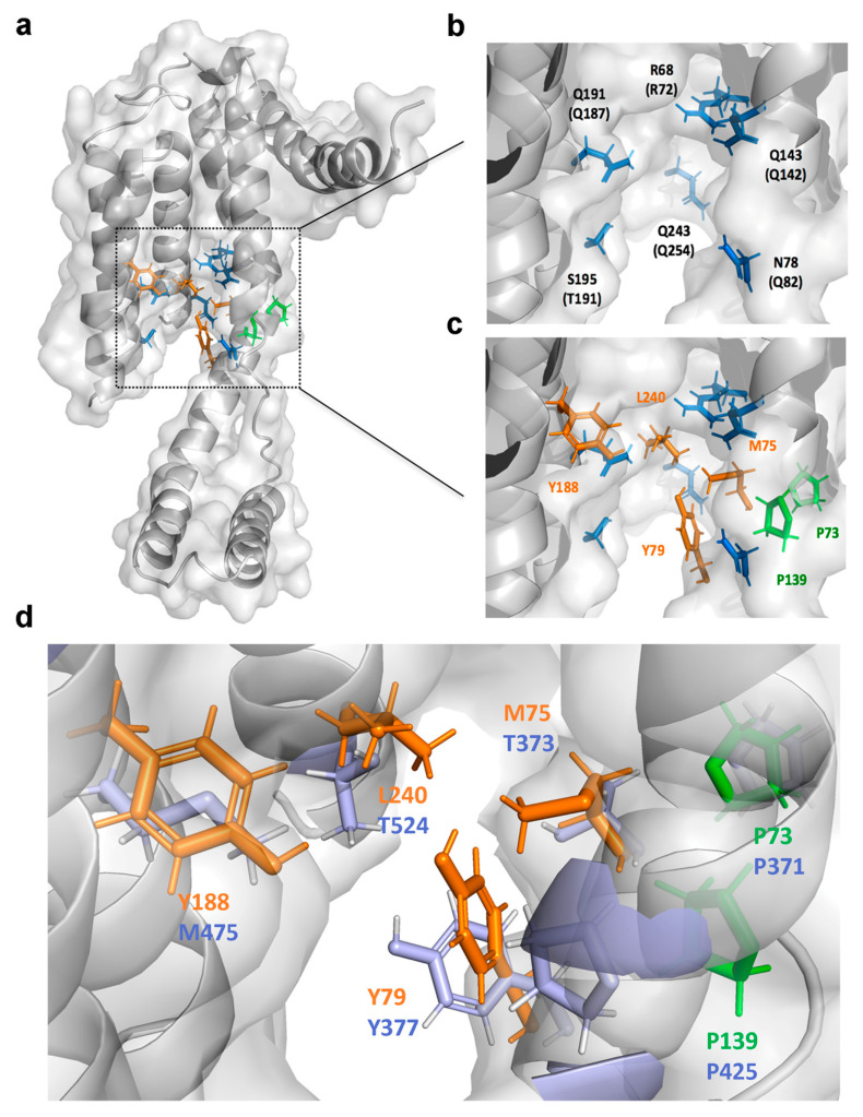
Substrate-contacting cavity of S. aureus YidC2. (a) The homology model of S. aureus YidC2 was built based on X-ray structures of B. halodurans YidC2 (PDB code: 3WO6 and 3WO7). (b) The hydrophilic groove of YidC is found to be conserved and the corresponding resides in B. halodurans YidCs are indicated in parentheses. (c) Mutations of YidC2 identified from drug-resistant S. aureus isolates are shown in green. The hydrophobic residues in the groove are shown in orange. (d) Comparison of Cpd36-contacting residues of YidC2 in S. aureus and E. coli shown in orange and purple, respectively.

