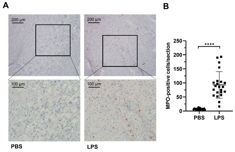
Figure 1.
Immunohistochemical analysis of MPO expression in hearts of PBS- and LPS-treated mice. C57BL/6 mice received a single i.p. injection of PBS (200 µL) or LPS in PBS (from Escherichia coli, 0111:B4 in PBS, 8 µg/g body weight) and were sacrificed 12 h after the injection. (A) Representative MPO-immunostainings of hearts isolated from PBS- and LPS-injected animals are shown at low and high magnification. (B) Statistical evaluation of MPO-positive cells in the sections of the hearts of PBS- or LPS-injected mice. Cryosections of eight different heart regions from PBS- or LPS-injected animals (n = 3) were counted manually for MPO-positive cells. Lines indicate mean ± SD values. Unpaired student’s t-test; **** p ≤ 0.0001.

