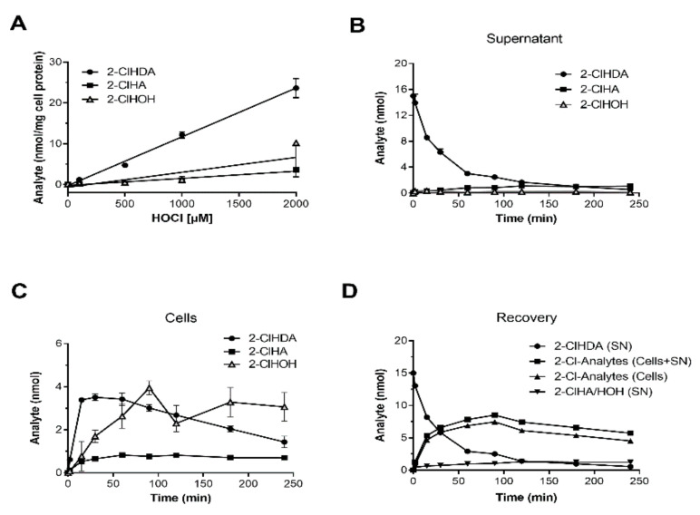
Figure 3.
In vitro formation and metabolism of 2-ClHDA in the murine HL-1 cardiomyocyte cell line. HL-1 cells were incubated with increasing concentrations of NaOCl or 2-ClHDA (15 µM). (A) After treatment with indicated concentrations of NaOCl for 1 h, cells were extracted in the presence of the corresponding internal standard as outlined in Materials and Methods. After conversion to their corresponding PFB-derivatives, 2-ClHDA, 2-ClHA, and 2-ClHOH concentrations were quantitated by NICI-GC–MS analysis. Results are displayed as mean ± SD (n = 3). (B,C) Cells were incubated with 15 µM 2-ClHDA for up to 4 h. At the indicated time points, 2-ClHDA, 2-ClHA, and 2-ClHOH concentrations were analyzed by NICI-GC–MS analysis in (B) the cellular supernatants and (C) HL-1 cells. Data represent mean ± SD values (n = 3). (D) Time-dependent recovery of 2-Cl-metabolites in HL-1 cells. Data represent loss of 2-ClHDA from the supernatant (SN), recovery of 2-Cl-Analytes (sum of 2-ClHDA, 2-ClHA, and 2-ClHOH in the SN or cells), and recovery of 2-ClHA plus 2-ClHOH in the supernatant (for reasons of clarity only mean values are shown).

