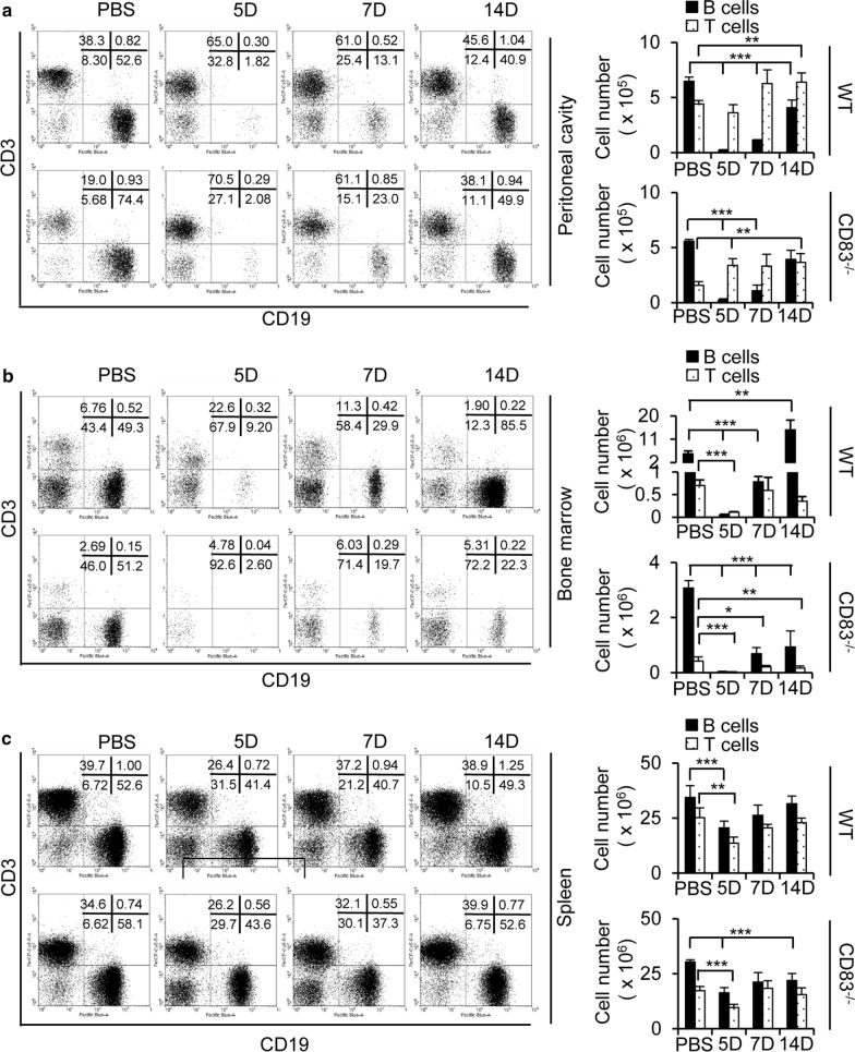
Fig. 2.
Population of lymphoid cells in the peritoneal cavity fluid, bone marrow, and spleen after intraperitoneal injection of A/WSN/1933 virus in C57BL/6J wild type mice and CD83 KO mice. a–c Wild type mice and CD83 KO mice (n = 5/group) were injected intraperitoneally with 5 × 10⁶ pfu of A/WSN/1933 virus. Peritoneal cells, bone marrow cells, and splenocytes were harvested at 5, 7, and 14 days after infection. Cells were enumerated and stained with fluorescence-conjugated antibodies to be analyzed by FACS. FSClowSSClow cells of peritoneal cavity fluid (a), bone marrow (b), and spleen (c) were sorted into CD19+ B cells and CD3+ T cells for all experimental groups of mice. The left panel displays the FACS profile with the percentage of the subpopulation in each quadrant. The right panel displays the absolute number of CD19+ B cells and CD3+ T cells in each subgroup. *P < 0.05, **P < 0.01, ***P < 0.001

