Figure 1.
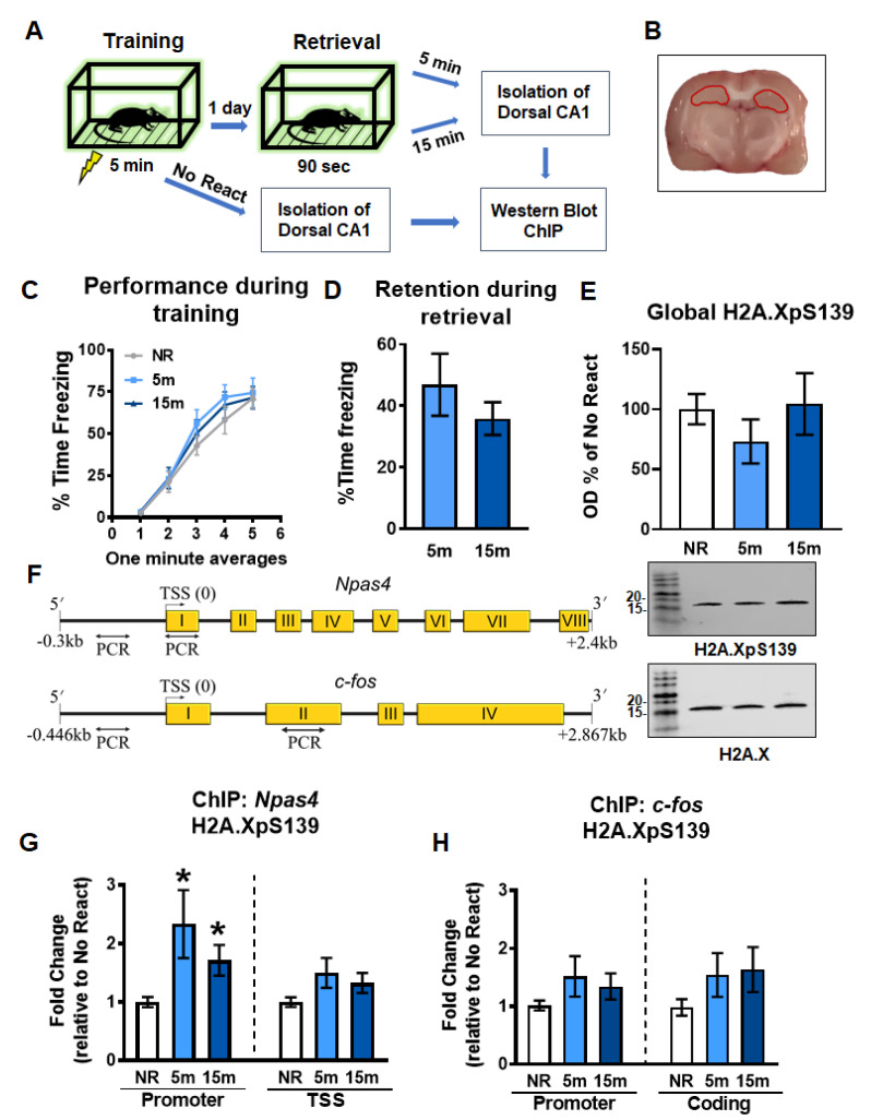
Gene-specific increases in H2A.XpS139 immediately after memory retrieval. (A) Experimental design. Rats were trained on a contextual fear conditioning task and exposed to the training context the following day to reactivate the memory. The CA1 region of the dorsal hippocampus was collected 5 or 15 min and processed for western blot and ChIP analyses. (B) Image depicting the dorsal hippocampus dissection, which is indicated in red. (C) Behavioral performance during the training session. (D) Memory retention during the retrieval session. (E) Western blot analysis revealed a moderate decrease in H2A.XpS139 levels in bulk histone extracts 5 min after the retrieval, which returned to baseline by 15 min (n = 12 per group). Representative bands show H2A.XpS139 (top) and total H2A.X (bottom) from the same gel. (F) Schematic showing primer targets for ChIP assays. The promoter and TSS regions of the Npas4 gene and the promoter and coding region of the c-fos gene were targeted. (G) ChIP analysis revealed an increase in H2A.XpS139 levels at the Npas4 promoter, but not TSS, region at 5 and 15 min following retrieval (n = 12 per group). (H) No changes in H2A.XpS139 were observed in either promoter or coding region of the c-fos gene following retrieval (n = 12 per group). NR: No React, TSS: Transcriptional start site. * Denotes p < 0.05 from No React.

