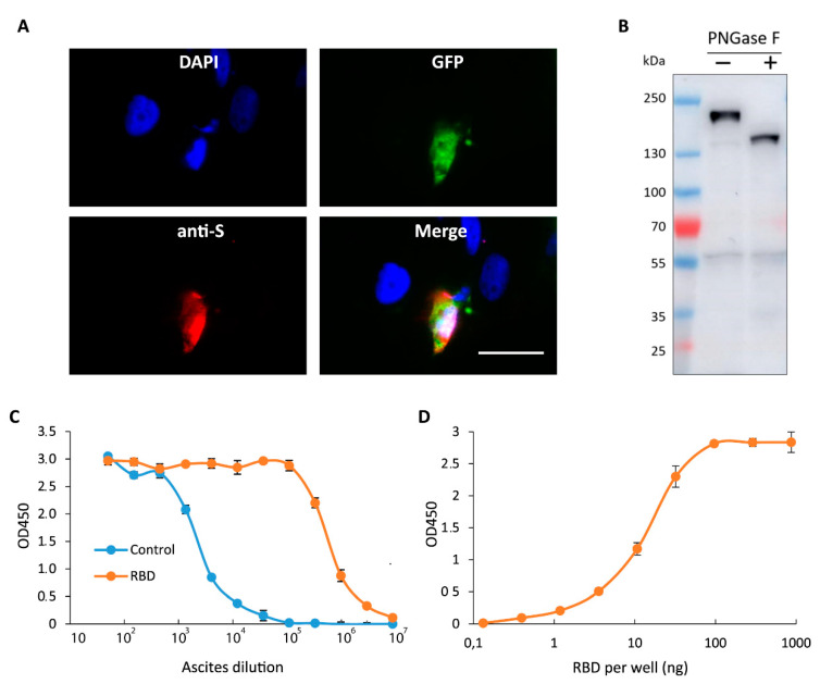
Figure 4.
Characterization of mAbs secreted by monoclone 11/9. (A) Fluorescence images of cells cotransfected with plasmids encoding GFP (green) and the full-length S protein and subsequently stained with DAPI (blue) and mAbs secreted by monoclone #11/9 (10 μg/mL; red). Scale bar = 20 μm, (B) Immunoblot analysis of cells transfected with plasmid encoding the full-length S protein and lysed by boiling in SDS. Lysate was incubated in a presence or absence of PNGase F. Membrane was stained with mAbs #11/9 (10 μg/mL). (C) Serial dilution ELISA showing reactivity of ascites formed by monoclone #11/9 against RBD (red) or control protein (blue) purified from E. coli. (D) ELISA showing reactivity of ascites (1:10,000 dilution) against different amounts of RBD purified from E. coli and immobilized in wells of EIA plate. Data are mean ± SD of three replicates.

