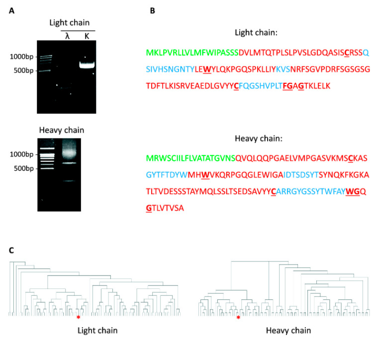
Figure 6.
Sequencing of anti-RBD SARS-CoV-2 mAbs. (A) PCR amplification of cDNA encoding immunoglobulins’ k-chain, λ-chain, and heavy-chain from monoclone #11/9. (B) Amino acid sequence of variable domains from light and heavy chains of 11/9 antibodies’. Different regions of immunoglobulins are highlighted: Ig leader sequence (green); framework regions (red); complementarity determining regions (blue); conserved amino acids (bold, underlined). (C) Dendrogram showing clustering of RBD binding antibodies based on the sequences of CDR3 in their light (left) and heavy (right) chains. mAbs #11/9 is indicated with red asterisk.

