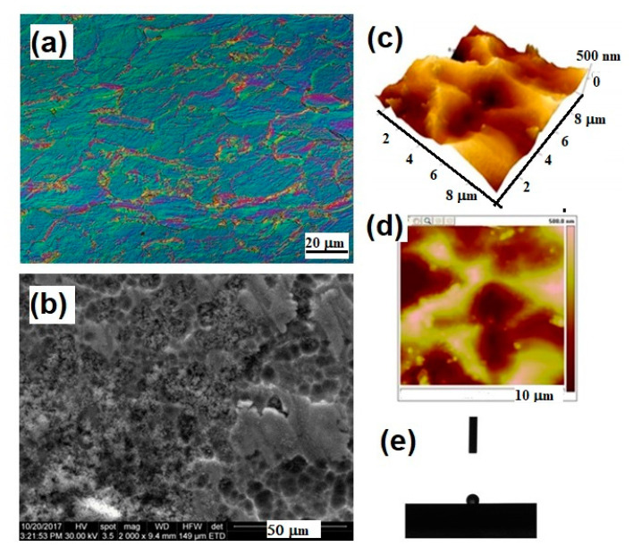
Figure 2.
Surface properties of Ti mesh for cranioplasty evidenced by different microscopic techniques: (a) light microscopy image in phase contrast, longitudinal section, 500×, Kroll reagent; (b) Scanning Electron Microscopy 2000×; (c,d) 3D and 2D Atomic Force Microscopy images; (e) contact angle investigation on the surface of the titanium mesh.

