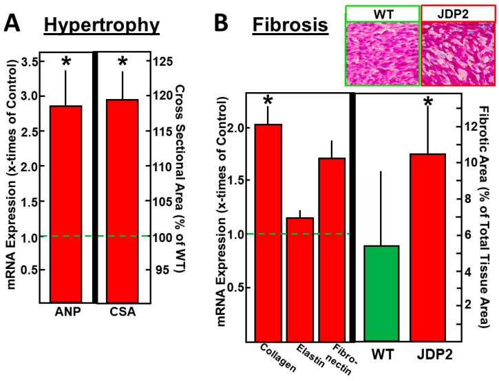
Figure 2.
Hypertrophy and fibrosis were analyzed in atria after five weeks of JDP2 overexpression. (A) mRNA expression of the hypertrophic marker gene ANP was determined by real-time RT-PCR and compared to 18SrRNA as a housekeeping gene (n = 11–12, p < 0.05 vs. WT) and a cross-sectional area of isolated cardiomyocytes was calculated from 30–40 cells per preparation (values are means ± SD of 4 independent culture preparations). (B) mRNA expression of fibrotic marker genes was determined by real-time RT-PCR and compared to 18SrRNA as a housekeeping gene (n = 11–12, p < 0.05 vs. WT), and fibrotic areas were determined after staining atrial tissue sections with azan dye (values are means ± SD of 5–6 tissue sections). * Difference from WT, p < 0.05.

