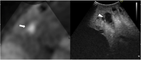
Figure 4.

Still intraoperative real-time virtual sonography image in Case 2. Preoperative diffusion-weighted magnetic resonance imaging (DW-MRI; left side) and CE-IOUS (right side) were synchronized. The CE-IOUS image demonstrated a hypo-echoic nodule (arrowhead) at the same site where DW-MRI demonstrated a lesion that was strongly suspected of being a liver metastasis (arrow).
