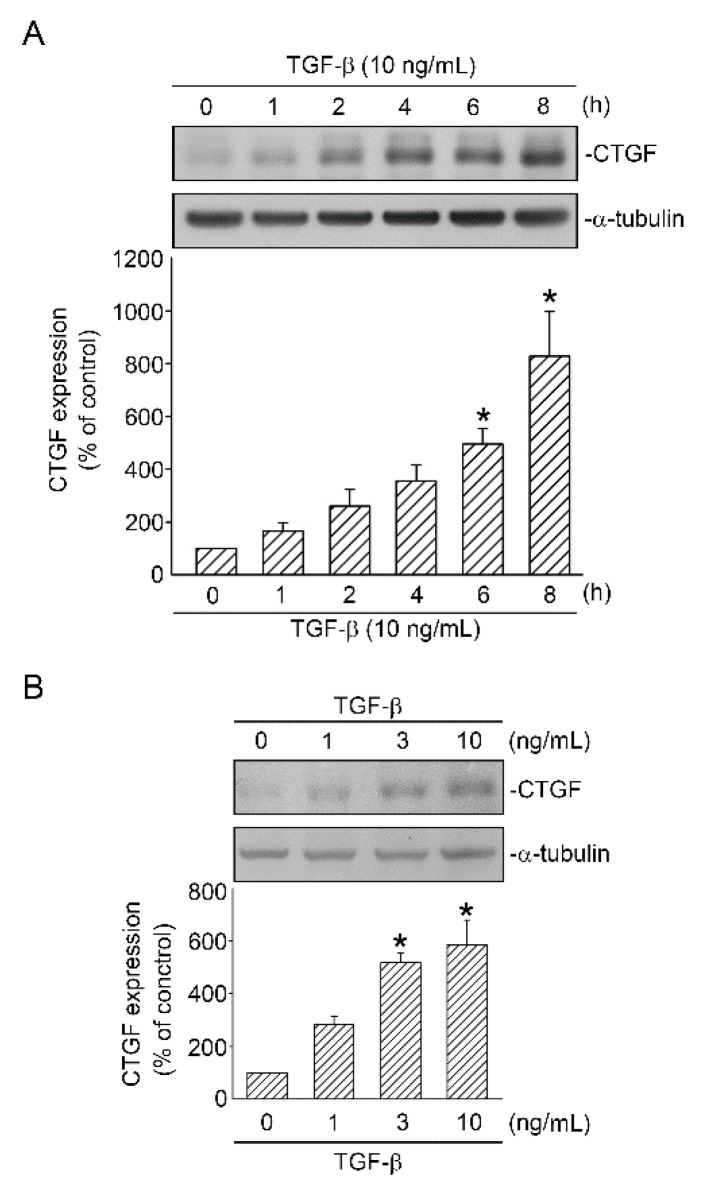
Figure 1.
TGF-β-induced CTGF expression in human lung epithelial A549 cells. (A) A549 cells were treated with TGF-β (10 ng/mL) for 0–8 h. Cell lysates were prepared, and CTGF and α-tubulin antibodies were detected through Western blotting. Data are expressed as mean ± SEM of three independent experiments. * p < 0.05, compared with the control group without TGF-β stimulation. (B) Cells were stimulated with TGF-β (0–10 ng/mL) for 6 h. Cell lysates were prepared, and CTGF and α-tubulin antibodies were detected through Western blotting. Data are presented as mean ± SEM of three independent experiments. * p < 0.05, compared with the control group without TGF-β treatment.

