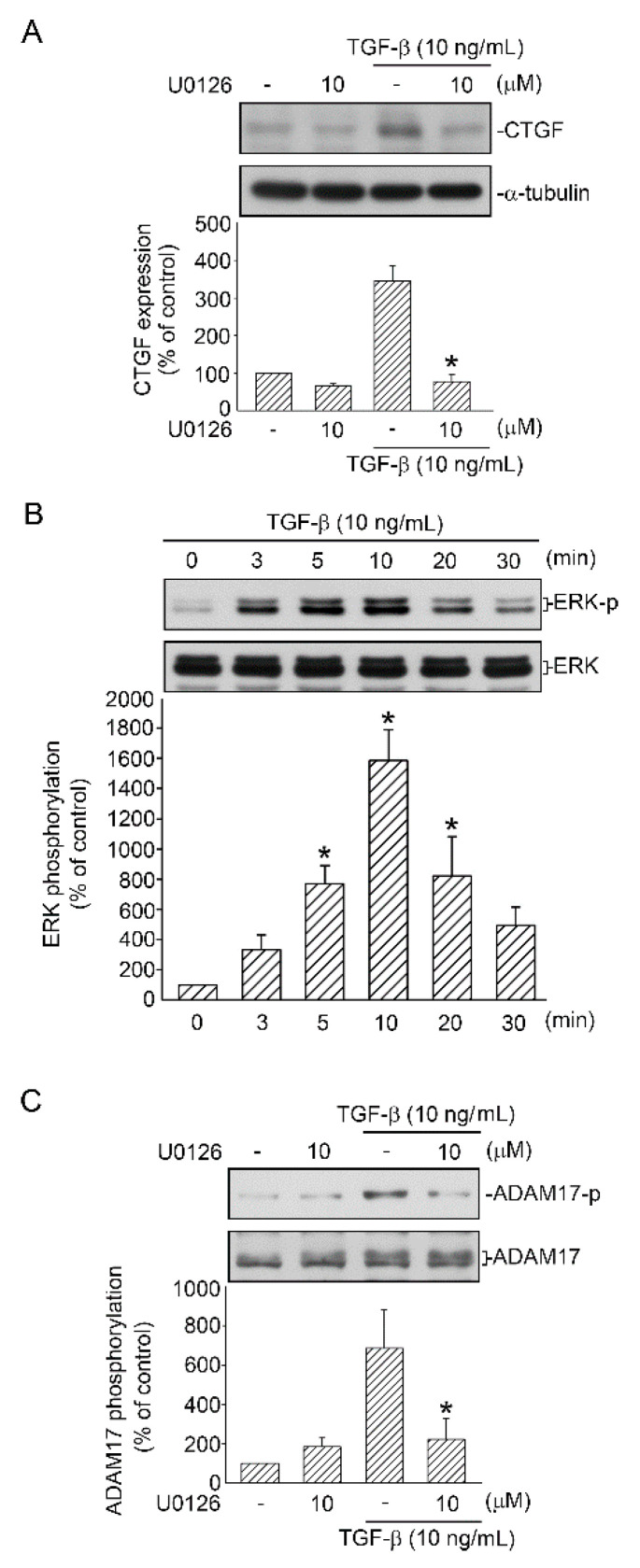
Figure 3.
Participation of ERK in TGF-β-induced CTGF expression and ADAM17 activation in human lung epithelial A549 cells. (A) Cells were processed with the ERK inhibitor U0126 (10 μM) for 20 min before they were stimulated with TGF-β (10 ng/mL) for an additional 6 h. CTGF or α-tubulin in cell lysates were immunodetected with specific antibodies. Data are expressed as mean ± SEM of three independent experiments. * p < 0.05, compared with the TGF-β group without U0126 treatment. (B) A549 cells were stimulated with TGF-β (10 ng/mL) for 0–30 min, and then the levels of ERK phosphorylation and ERK in cell lysates were immunodetected with specific antibodies. Data are presented as mean ± SEM of four independent experiments. * p < 0.05, compared with the control group without TGF-β treatment. (C) Cells were treated with the ERK inhibitor U0126 (10 μM) for 20 min before they were stimulated with TGF-β (10 ng/mL) for an additional 10 min. Cell lysates were prepared, and specific antibodies for ADAM17 phosphorylation and ADAM17 were detected through Western blotting. Data are presented as mean ± SEM of three independent experiments. * p < 0.05, compared with the TGF-β group without U0126 treatment.

