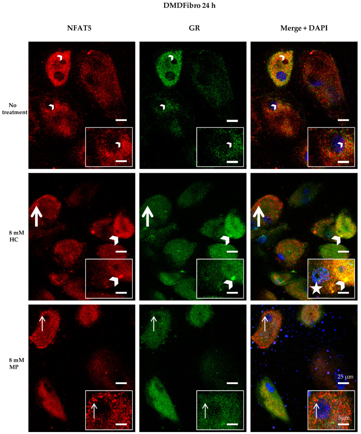
Figure 4.
NFAT5 and GR localization in DMD fibroblasts treated for 24 h with hydrocortisone or methylprednisolone NFAT5 (red) and GR (green) are visualized by immunofluorescence. Nuclei are stained in DAPI (blue). Small yellow dots are seen in untreated DMDFibro (small white arrowheads). Here, both NFAT5 and GR are superimposed. Large yellow colocalization is only seen in DMDFibro with 8 mM HC (large white arrowheads). Orange dots are visible both after 8 mM HC and 8 mM MP treatment for 24 h, with small orange in 8 mM (small white arrow) and large ones in 8 mM HC (large white arrow). In orange dots, NFAT5 and GR are very close to each other but not superimposed. After 24 h with 8 mM HC, DMDFibro display very small yellow structures in the nucleus (white star), which stain for NFAT5 and GR (insets; n = 3).

