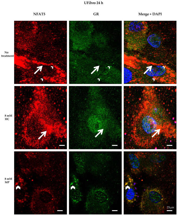
Figure 5.
NFAT5 and GR localization in unaffected skeletal muscle fibroblasts treated for 24 h with hydrocortisone or methylprednisolone NFAT5 (red) and GR (green) are visualized by immunofluorescence. Nuclei are stained in DAPI (blue). Small yellow dots (small white arrowheads) and large orange dots (large white arrows) are seen in untreated UFibro or after treatment for 24 h with 8 mM HC. After 24 h with 8 mM MP, UFibro display large amounts of yellow round structures in the perinuclear area (large white arrowheads), which stain for NFAT5 and GR (n = 3).

