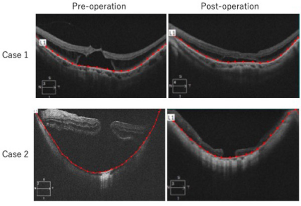
Figure 3.

Flattened curvature of posterior fundus after lamellar scleral resection and infolding of the remaining sclera in patients with myopic retinoschisis and macular hole retinal detachment. Pre- and post-operative OCT in case 1 and 2 are shown. Posterior segment distance defined as the distance of retinal pigment epithelial line (red) using photo analyzer (AreaQ, Japan). Shorter posterior segment distance suggests flattened curvature of the posterior fundus after surgery.
