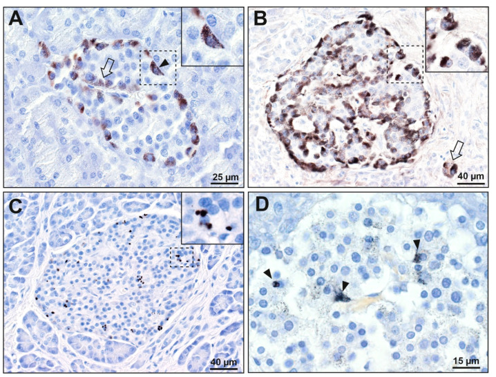
Figure 2.
Mercury in pancreatic islet cells. (A–C): pancreatectomy samples. (D): autopsy sample. The areas in the dashed rectangles are shown at higher magnifications in the insets. (A) Black mercury grains are present in most peripheral cells (e.g., arrowhead) of this islet, as well as in a few internal cells adjacent to microvessels (e.g., arrow). (B) Black mercury grains in this islet are present in the cytoplasm of all peripheral cells, as well as in most internal cells adjacent to microvessels. Two cells outside the islet (arrow, probably ectopic islet cells) contain mercury. (C) Numerous dense black mercury granules are visible within peripheral and internal cells of this islet, usually adjacent to cell nuclei. (D) Fine black mercury grains are present in many scattered cells of this islet, with denser staining in three cells (arrowheads). Autometallography/hematoxylin.

