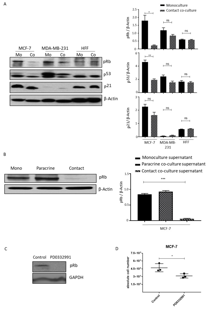
Figure 6.
Contact lipoaspirate culture inhibits MCF-7 proliferation via the retinoblastoma-mediated pathway. (A) Cell lysates from MCF-7, MDA-MB-231 or HFF contact-co-cultured with lipo-aspirates were collected in Ripa lysate buffer. The phosphorylation of retinoblastoma protein (Rb) and the expression of p53 and p21 were analyzed by western blotting using specific antibodies. β-Actin was employed as the loading control. Fold changes in densitometric band intensities, acquired by image J and normalized to β-Actin, were compared and plotted. (B) Cell lysates from MCF-7 cells cultured in media from either monocultured, paracrine-cultured or contact-cultured MCF-cells with lipo-aspirates were collected in Ripa buffer and blotted for phosphorylated retinoblastoma protein (Rb). β-Actin was employed as the loading control. (C,D) MCF-7 cells were incubated with 10µM PD0332991 and cell lysates were blotted for Rb protein phosphorylation (C) and counted at day 4 post seeding (D). p value < 0.05 = *, p < 0.01 = **, p < 0.001 = ***, ns = non-significant.

