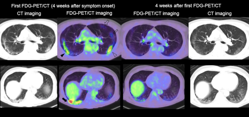
Figure 3.

FDG-PET/CT imaging of lung lesion in COVID-19. Left side: CT and fused FDG-PET/CT image of the chest in the first examination (4 weeks after symptom onset). Right side: CT and fused FDG-PET/CT image of the chest (4 weeks after the first FDG-PET/CT examination). Intense FDG uptake was seen in liner opacity (black arrowhead), reticular opacity with consolidation (black arrow), and grand glass opacity with consolidation (open arrowhead) in the lung. Moderate FDG uptake was confirmed in grand glass opacity (open arrows) at the left upper lobe. All the FDG uptake in the first examination was significantly decreased in the second PET/CT scan.
