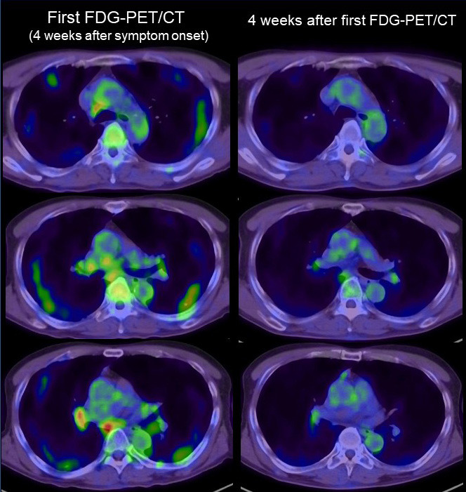
Figure 4.

FDG-PET/CT imaging of lymph nodes in COVID-19. Left side: FDG-PET/CT image of the chest in the first examination (4 weeks after symptom onset). Right side: FDG-PET/CT image of the chest (4 weeks after the first FDG-PET/CT examination). Intense FDG uptake was seen in mediastinal and hilar lymph nodes. All the FDG uptake in the first examination was significantly decreased in the second FDG-PET/CT scan.
