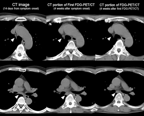
Figure 5.

Change of CT feature of lymph node over time. Left side: CT image (14 days from symptom onset), Middle: CT portion of FDG-PET/CT (4 weeks after symptom onset), Right side: CT portion of second FDG-PET/CT (4 weeks after first FDG-PET/CT examination). CT image (14 days from symptom onset) showed no evidence of mediastinal lymph node swelling (arrowhead). Although the size of the lymph node is not significant, it was increased compared to 2 weeks from CT imaging and decreased within 4 weeks after the first FDG-PET/CT examination.
