Fig. 1.
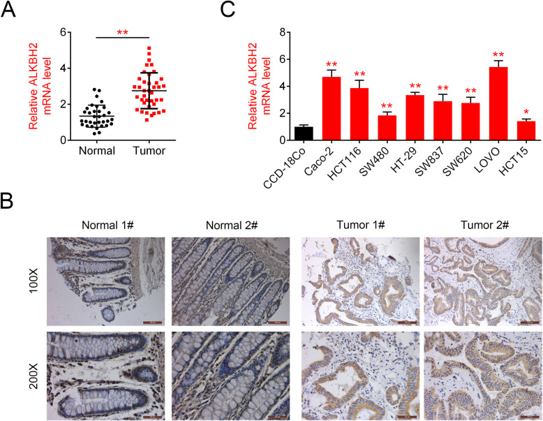
ALKBH2 is upregulated in CRC tissues and cells. a Relative ALKBH2 expression in CRC tissues (N = 39) and paracarcinoma (N = 33), quantified using qRT-PCR assays, **P < 0.01 vs. paracarcinoma. The mRNA levels of ALKBH2 are significantly increased in CRC tissues compared to that of control. b Representative IHC images showing ALKBH2 staining in CRC and paracarcinoma, **P < 0.01 vs. paracarcinoma. The percentage of ALKBH2-positive cells is higher in CRC tissues than para-carcinoma. c ALKBH2 expression in CRC lines (Caco-2, HCT116, SW480, HT-29, SW837, SW620, LOVO, and HCT15) compared to the colorectal cell (GES-1) as quantified using qRT-PCR assays. ALKBH2 is highly expressed in all colorectal carcinoma cells compared with normal colorectal epithelial cells

