
Fig. 1.
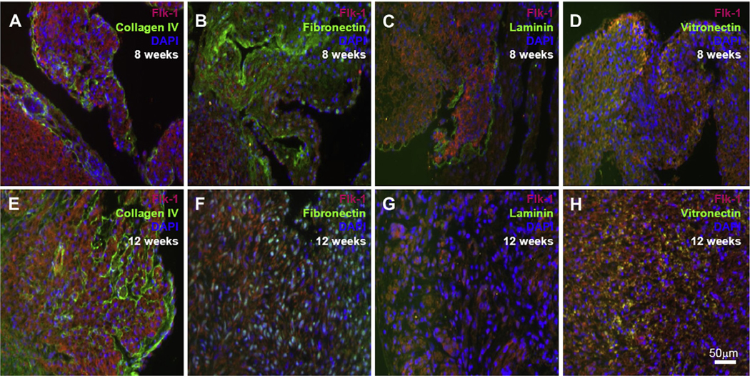
Immunofluorescence staining of first-trimester human heart tissues shows the endogenous Flk-1 expressing CPC (red) as well as the expression of ColIV (A, E), fibronectin (B, F), laminin (C, G) and vitronectin (D, H) all in green. Cell nuclei are identified with DAPI staining (blue). Scale bars 50 μm.