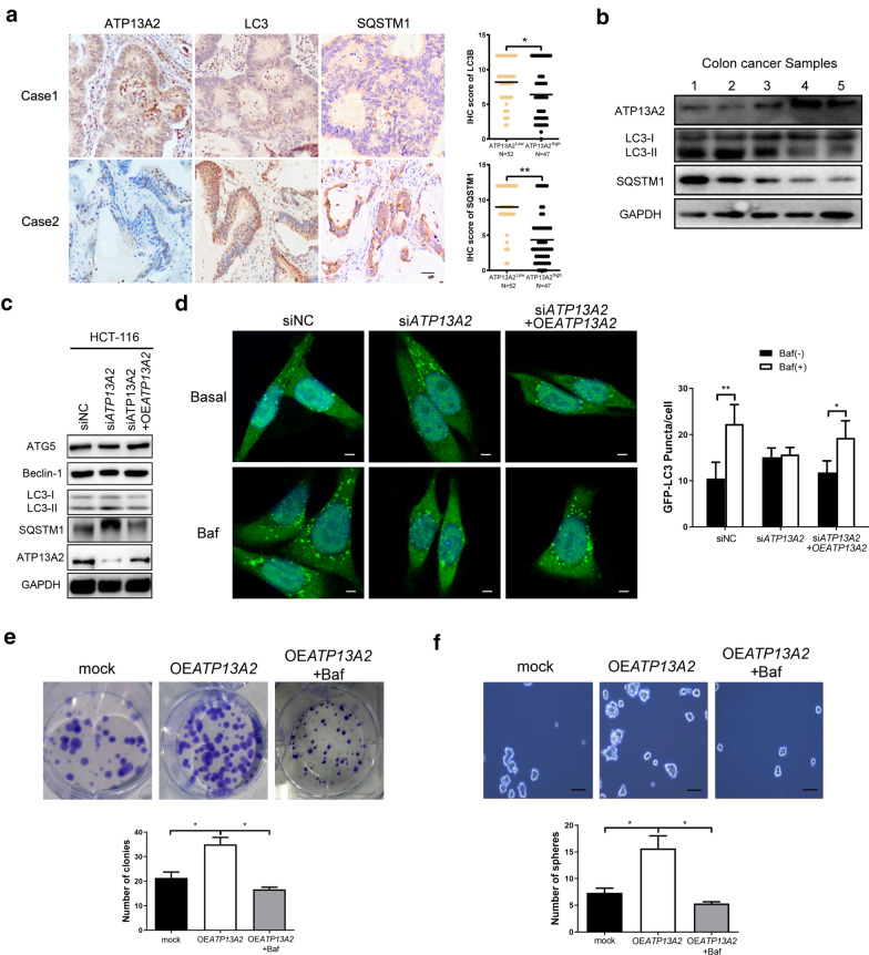
Fig. 5.
Involvement of ATP13A2 in the stemness of colon cancer cells through regulation of autophagy. a Representative immunohistochemical staining of ATP13A2, LC3, SQSTM1 (Left panel) and statistical analysis (Right panel) in colon cancer tissues (n = 99). Scale bar = 50 µm. (b) ATP13A2, LC3 and SQSTM1 were detected by western blotting (WB) in five fresh human colon cancer samples. c The protein expression of ATP13A2, SQSTM1, LC3, Beclin-1, and ATG5 in ATP13A2-knockdown HCT-116 cell groups, in ATP13A2-knockdown groups rescued by ATP13A2 overexpression, and in control cells. d The representative image (Left panel) and number (Right panel) of green puncta (LC3 protein) from HCT-116-siNC and HCT-116-siATP13A2 treated with and without bafilomycin; HCT-116 cells were co-transfected with indicated siRNA and GFP-LC3 plasmid. Some siATP13A2 cultures were rescued by the overexpression of ATP13A2 (OEATP13A2). After 48 h, cultures were treated with dimethyl sulfoxide (DMSO) or bafilomycin (5 nM) for 2 h. Scale bar = 5 μm. N = 5, with 5–10 cells imaged per experiment, values are means ± SD. e–f The number of colonies (e) and spheres (f) in the mock group, OEATP13A2 group, and OEATP13A2 + bafilomycin (5 nM) group. *P < 0.05; **P < 0.01;

