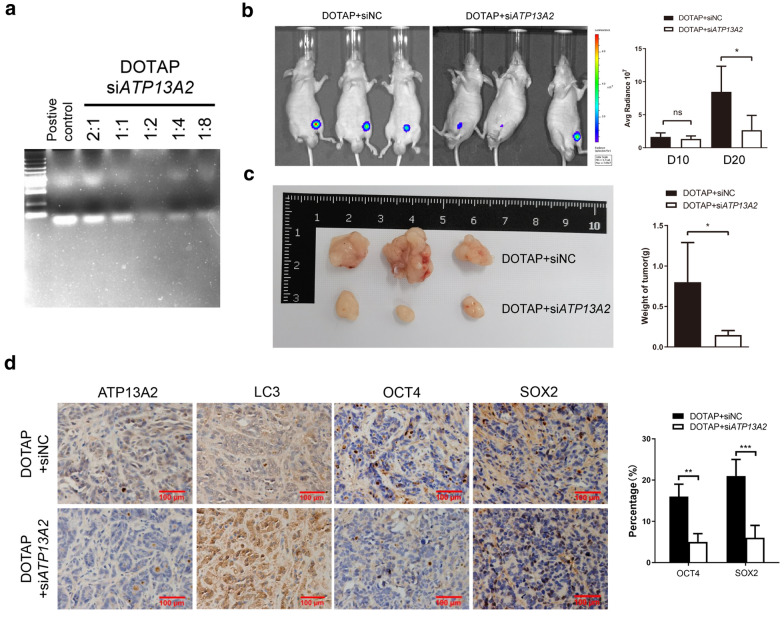
Fig. 6.
Inhibition of xenograft tumor proliferation by in vivo treatment with ATP13A2 siRNA. a Gel retardation assay shows the effects of using different ratios of DOTAP and siATP13A2. b Nude mice (n = 3) were implanted with SW480 cells, and after 10 days, the mice were treated with siNC or siATP13A2 mixed with DOTAP. Bioluminescence (Left panel) and qualification (Right panel) images of tumors in these mice at 20 day after incubation were shown. c Representative image and quantitative analysis of weight of the xenograft tumor were shown when mice were sacrificed. d Representative images (Left panel) and quantification (Right panel) of SOX2, OCT4, LC3, and ATP13A2 in xenograft tumors treated with siNC or siATP13A2 mixed with DOTAP (n = 3). *, P < 0.05; **, P < 0.01; ***, P < 0.01; ns, no significant

