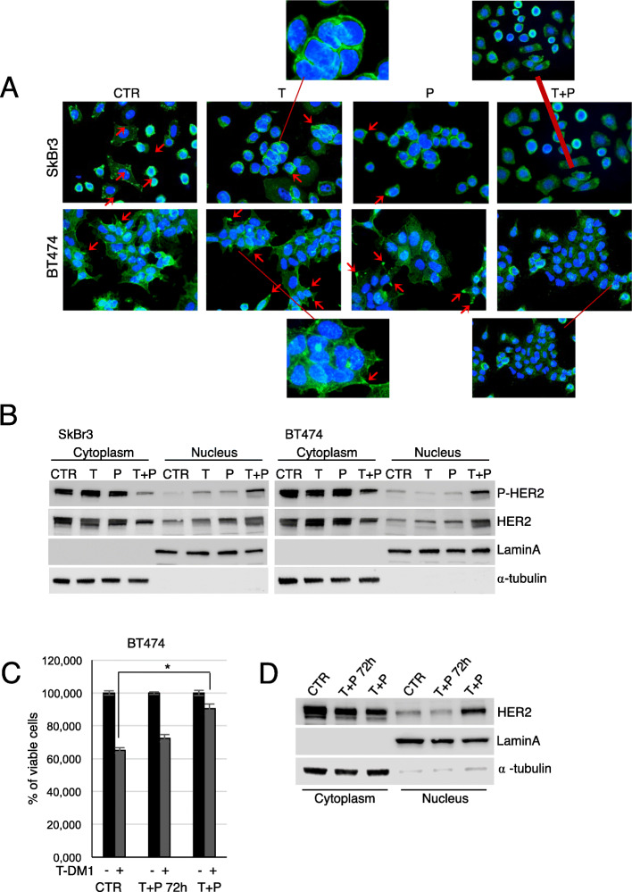
Fig. 3.
Prolonged trastuzumab+pertuzumab induces HER2 nuclear translocation. Control, T, P, and T + P cell lines were plated on poly-l lysine coated slides, and stained 24 hous later with anti-HER2 (green signal) (a). These cells were counterstained with Hoechst to highlight nuclei. Red arrows indicate HER2 localization on cellular protrusions. Cytoplasmic and nuclear fractions extracted from control, T, P, and T + P cells were analysed by Western Blot (WB) for the expression of phosphorylated and total HER2. Lamin A and α-tubulin were used to validate purity of nuclear and cytoplasmic extracts respectively (b). Following pre-treatment with 5 μg/ml trastuzumab + 5 μg/ml pertuzumab for 72 h, cell viability of control cells, pre-treated and T + P BT474 exposed to1 μg/ml T-DM1 for 72 h was evaluated by Crystal Violet Assay (c). Cytoplasmicand nuclear fractions of control, pre-treated and T + P BT474 cells were analysed by WB for the expression of total HER2 (d)

