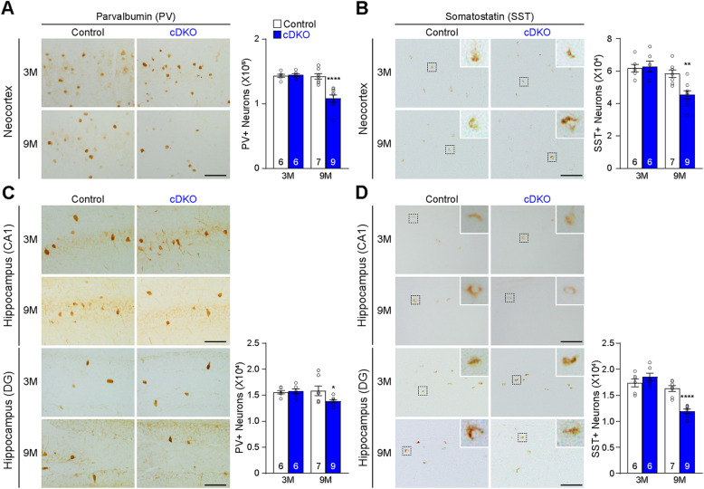
Fig. 4.
Progressive decreases of PV+ and SST+ interneurons in the cerebral cortex of IN-PS cDKO mice. a Left: PV immunostaining of comparable sagittal sections in the neocortex of IN-PS cDKO and littermate control brains at 3 and 9 months of age. Right: Stereological quantification shows age-dependent decreases in the number of PV-immunoreactive interneurons in the neocortex of IN-PS cDKO mice compared to that in control mice (F1, 24 = 16.20, p = 0.0005; 3 M: Control 1.43 ± 0.02 × 10⁵, cDKO 1.45 ± 0.02 × 10⁵, p > 0.99; 9 M: Control 1.43 ± 0.04 × 10⁵, cDKO 1.09 ± 0.05 × 10⁵, p < 0.0001, two-way ANOVA with Bonferroni’s post hoc comparisons). b Left: SST immunostaining of comparable sagittal sections in the neocortex of IN-PS cDKO and littermate control brains at 3 and 9 months of age. Inserts show higher power views of the boxed areas. Right: Stereological quantification shows age-dependent decreases in the number of SST-immunoreactive interneurons in the neocortex of IN-PS cDKO mice compared to that in control mice (F1, 24 = 6.61, p = 0.0168; 3 M: Control 6.17 ± 0.24 × 10⁴, cDKO: 6.28 ± 0.34 × 10⁴, p > 0.99; 9 M: Control 5.84 ± 0.24 × 10⁴, cDKO 4.55 ± 0.24 × 10⁴, p = 0.0027, two-way ANOVA with Bonferroni’s post hoc comparisons). c Left: PV immunostaining of comparable sagittal sections in hippocampus area CA1 (CA1) and the dentate gyrus (DG) of IN-PS cDKO and littermate control brains at 3 and 9 months of age. Right: Stereological quantification shows age-dependent decreases in the number of PV-immunoreactive interneurons in the entire hippocampus of IN-PS cDKO mice compared to that in control mice (F1, 24 = 3.84, p = 0.062; 3 M: Control 1.56 ± 0.03 × 10⁴, cDKO 1.58 ± 0.04 × 10⁴, p > 0.99; 9 M: Control 1.59 ± 0.10 × 10⁴, cDKO 1.38 ± 0.03 × 10⁴, p = 0.0244, two-way ANOVA with Bonferroni’s post hoc comparisons). d Left: SST immunostaining of comparable sagittal sections in hippocampus area CA1 (CA1) and the dentate gyrus (DG) of IN-PS cDKO and littermate control brains at 3 and 9 months of age. Inserts show higher power views of the boxed areas. Right: Stereological quantification shows age-dependent decreases in the number of SST-immunoreactive interneurons in the entire hippocampus of IN-PS cDKO mice compared to that in control mice (F1, 24 = 20.92, p = 0.0001; 3 M: Control 1.74 ± 0.08 × 10⁴, cDKO 1.85 ± 0.08 × 10⁴, p = 0.44; 9 M: Control 1.63 ± 0.06 × 10⁴, cDKO 1.19 ± 0.04 × 10⁴, p < 0.0001, two-way ANOVA with Bonferroni’s post hoc comparisons). Scale bar: 100 μm. All data represent mean ± SEM. *p < 0.05, **p < 0.01, ****p < 0.0001. The value in the column indicates the number of mice used in each experiment

