Figure 1.

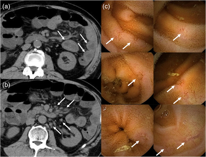
Computed tomography shows diffuse wall thickness of the jejunum (a, arrow) and lymph node swellings of the mesentery (b, arrow). Capsule endoscopy shows multiple erosions or small ulcers in the small intestine (c, arrow).

Computed tomography shows diffuse wall thickness of the jejunum (a, arrow) and lymph node swellings of the mesentery (b, arrow). Capsule endoscopy shows multiple erosions or small ulcers in the small intestine (c, arrow).