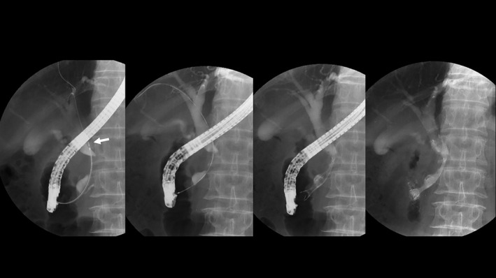
Figure 2.

Half‐covered metallic stent placement. Endoscopic retrograde cholangiography and intraductal ultrasonography confirmed the position of the cystic duct confluence. At the second endoscopic retrograde cholangiography, the position of cystic duct confluence (arrow), the length of biliary stricture, and the distance from the papilla Vateri to the upper site of the stricture were determined using a measuring guide wire. The half‐covered stent was selected and deployed.
