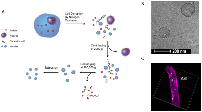
Figure 8:
(A) The preparation of cell membrane-derived nanovesicles via nitrogen cavitation and a series of centrifugations. After purification, the intracellular components are removed and purified nanovesicles are obtained. (B) Cryo-TEM image of HL-60 cell membrane-formed nanovesicles. (C) The intravital image shows a cremaster venule from a live mouse following the i.v. injection of DiO fluorescently labeled nanovesicles (green) and Alex-Fluor-647 anti-CD31 (pink) to label the blood vessel. Copyright 2016 Elsevier.

