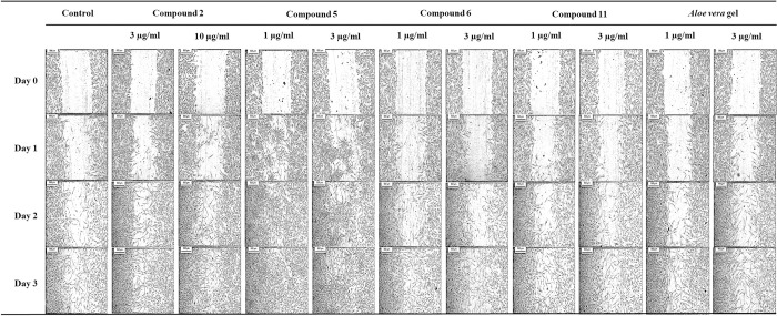
Fig 3. Effect of compounds 2, 5, 6, 11 and A. vera gel on HDF migration.
Images were captured at day 0 and showed that an artificial wounded monolayer was created using the scratch assay. Then treated with 3 and 10 μM 15,16-epoxy-6α-O-acetyl-8β-hydroxy-19-nor-clero-13(16),14-diene-17,12,18,2-diolide (2), 1 and 3 μM (+)-catechin (5), 1 and 3 μM quercetin (6), 1 and 3 μM myricetin (11), 1 and 3 μg/ml A. vera gel and control without treatment. Another set of images of fibroblast cell migration were captured at day 1, 2 and 3 after incubation. The cleared areas represented wound and shaded areas resulting in cell migration, which represents the wound closure.

