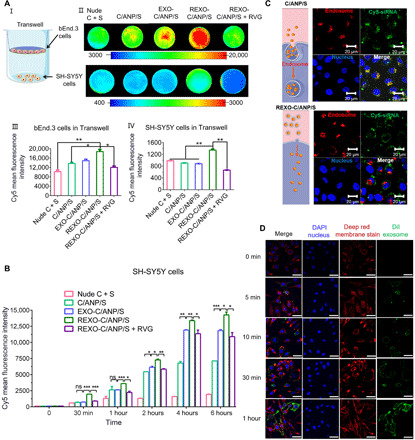
Fig. 2. In vitro drug delivery.

(A) NP internalization in Transwell cells in 12 hours. I: Scheme of Transwell instrument. II: Cy5-siRNA internalization of bEnd.3 cells (top) and the SH-SH5Y cells (bottom). III: Cy5 mean fluorescence intensity in NP-treated bEnd.3 cells in Transwell model. IV: Cy5 mean fluorescence intensity in NP-treated SH-SH5Y cells in the Transwell model. (B) Cy5 mean fluorescence intensity detected by flow cytometry in SH-SH5Y cells after NP incubation in 0 min, 30 min, 1 hour, 2 hours, 4 hours, and 6 hours. ns, not significant. (C) Assessment by CLSM of SH-SY5Y cells after NP incubation in 4 hours. Endosome was labeled with LysoTracker red. Cy5-siSNCA, green. (D) Assessment by CLSM of SH-SY5Y cells after NP incubation in 0 min, 5 min, 10 min, 30 min, and 1 hour. Cell membrane was labeled with CellMask deep red membrane stain, and exosome was labeled with DiI.*P < 0.05, **P < 0.01, and ***P < 0.001. DAPI, 4′,6-diamidino-2-phenylindole.
