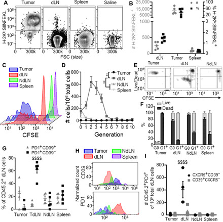
Fig. 4. The TdLN is a niche that supports Ag-specific CD8+ T cell priming and survival.

SIINFEKL presentation (measured by 25D1.16 staining for H-2Kb:SIINFEKL) by CD45+ cells (A) and cDCs (B) of the tumor, dLN, and spleen in B16F10-OVA tumor–bearing animals. Representative carboxyfluorescein diacetate succinimidyl ester (CFSE) dilution histograms (C) showing proliferation by tumor Ag–specific CD45.2+ donor cells in the tumor, dLN, NdLN, and spleen 72 hours after transfer into day 7 tumor-bearing CD45.1 animals, as quantified in (D). Representative flow cytometry plots (E) and quantification (F) of proliferative generation (CFSE signal) and viability tumor Ag–specific donor cells in the tumor, dLN, and NdLN. (G to I) Phenotype of tumor Ag–specific donor cells in the tumor, dLN, NdLN, and spleen 72 hours after transfer into day 7 tumor-bearing CD45.1 mice. (G and H) CD44, CD39, and PD1 expression quantification (G) and representative histograms (H). (I) CXCR5 and CD39 expression quantification. * indicates significance by two-way ANOVA with Tukey’s comparison (** indicates P < 0.01); $ indicates significance relative to all other groups by two-way ANOVA with Tukey’s comparison ($$$$ indicates P < 0.0001); n = 5 to 8 animals; (A) to (I) are representative of two experiments.
