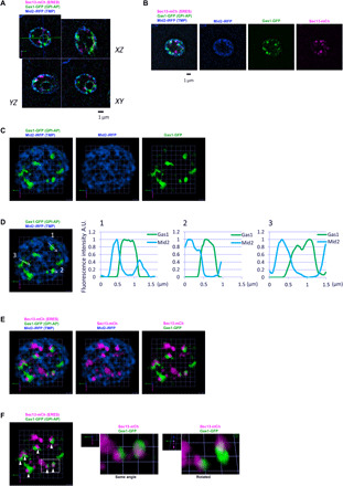
Fig. 1. Newly synthesized C26 ceramide–based GPI-AP cargos form clusters in the ER membrane adjacent to specific ERES.

sec31-1 cells expressing galactose-inducible secretory cargos, the very long acyl chain (C26) ceramide GPI-AP Gas1-GFP (GPI-AP, green) and the transmembrane protein Mid2-iRFP (TMP, blue), and constitutive ERES marker Sec13-mCherry (ERES, magenta) were incubated at 37°C for 30 min, shifted down to 24°C and imaged by SCLIM after 5 min. (A to C) Representative merged or individual 2D images of one plane (A), 2D projection images of 10 z-sections (B), or 3D cell hemisphere images (C) of cargo and ERES markers are shown. Scale bar, 1 μm (A and B). Scale unit, 0.551 μm (C). Gas1-GFP was detected in discrete ER zones or clusters, whereas Mid2-iRFP was detected and distributed throughout the ER membrane (C). (D) Graphs show relative fluorescence intensities of Gas1-GFP and Mid2-iRFP along the white arrow lines in the Gas1-GFP clusters (left). A.U., arbitrary units. (E and F) Representative merged 3D images of cargo and ERES markers. Gas1-GFP clusters were detected adjacent to specific ERES. Scale unit, 0.551 μm. (F) The white filled arrowheads mark Gas1-GFP clusters associated with ERES. Middle and right panels show a merged enlarged 3D image and rotated view of selected Gas1-GFP clusters.
