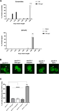
Fig. 3. ER clustering of C26 ceramide–based GPI-APs requires the presence of C26 ceramides in the membrane and interaction with the p24 complex receptor through the GPI-glycan.

(A) The cellular membranes of GhLag1 mainly contain shorter C18-C16 ceramide, while the GPI anchor of Gas1-GFP still has C26 IPC as in wild-type cells. Upper graph: acyl chain length analysis by mass spectrometry (MS) of ceramides from cellular membranes of wild-type (Wt) and GhLag1p strains. Data represent the percentage of total ceramides. Mean of three independent experiments. Error bars = SD. Two-tailed, unpaired t test. ****P < 0.0001. Lower graph: acyl chain length analysis by MS of IPCs present in the GPI anchor of Gas1-GFP (GPI-IPC) expressed in wild-type and GhLag1p strains. Data represent the percentage of total IPC signals. Mean of five independent experiments. Error bars = SD. Two-tailed, unpaired t test. ns, not significant. P = 0.9134. (B) Fluorescent micrographs of sec31-1, sec31-1 GhLag1, sec31-1 emp24Δ, sec31-1 ted1Δ, and sec31-1 lst1Δ cells expressing galactose-inducible Gas1-GFP were incubated at 37°C for 30 min and visualized by conventional fluorescence microscopy after shifting down to 24°C. White arrowheads: ER Gas1-GFP clusters. Open arrowheads: unclustered Gas1-GFP distributed throughout the ER membrane showing the ER-characteristic nuclear ring staining. Scale bar, 5 μm. (C) Quantification of micrographs described in (B). Average percentage of the cells with dot-like Gas1-GFP structures. n ≥ 300 cells in three independent experiments. Error bars = SD. Two-tailed, unpaired t test. ****P < 0.0001.
