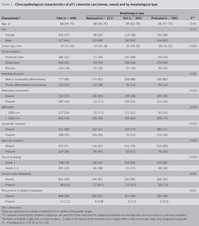
Table 1.
Clinicopathological characteristics of pT1 colorectal carcinomas, overall and by morphological type
| Characteristica | Total (n = 909) | Morphological type | Pb | ||
| Depressed (n = 211) | Flat (n = 304) | Protruded (n = 394) | |||
| Age, yr | 68 (59–75) | 68 (60–75) | 69 (62–76) | 66 (57–73) | 0.49 |
| Sex | 0.14 | ||||
| Female | 332 (37) | 68 (32) | 114 (38) | 150 (38) | |
| Male | 577 (64) | 143 (68) | 190 (62) | 244 (62) | |
| Tumor size, mm | 19 (13–25) | 14 (10–18) | 25 (18–35) | 18 (13–23) | <0.001 |
| Tumor location | |||||
| Proximal colon | 282 (31) | 71 (34) | 147 (48) | 64 (16) | |
| Distal colon | 463 (51) | 93 (44) | 100 (33) | 270 (69) | |
| Rectum | 164 (18) | 47 (22) | 57 (19) | 60 (15) | |
| Histological grade | 0.18 | ||||
| Well or moderately differentiated | 777 (85) | 174 (82) | 268 (88) | 335 (85) | |
| Poorly differentiated or mucinous | 132 (15) | 37 (18) | 36 (12) | 59 (15) | |
| Adenoma component | <0.001 | ||||
| Absent | 522 (57) | 196 (93) | 145 (48) | 181 (45) | |
| Present | 387 (43) | 15 (7.1) | 159 (52) | 213 (54) | |
| SM depth | <0.001 | ||||
| <1000 μm | 217 (24) | 15 (7.1) | 111 (37) | 91 (23) | |
| ≥1000 μm | 692 (76) | 196 (93) | 193 (63) | 303 (77) | |
| Lymphatic invasion | <0.001 | ||||
| Absent | 621 (68) | 107 (51) | 234 (77) | 280 (71) | |
| Present | 288 (32) | 104 (49) | 70 (23) | 114 (29) | |
| Vascular invasion | <0.001 | ||||
| Absent | 672 (7) | 116 (55) | 241 (79) | 315 (80) | |
| Present | 237 (26) | 95 (45) | 63 (21) | 79 (20) | |
| Tumor buddingc | <0.001 | ||||
| Grade 1 | 708 (78) | 130 (62) | 253 (83) | 325 (83) | |
| Grade 2–3 | 201 (22) | 81 (38) | 51 (17) | 69 (18) | |
| Lymph node metastasis | 0.08 | ||||
| Absent | 851 (94) | 194 (92) | 292 (96) | 365 (93) | |
| Present | 58 (6.4) | 17 (8.1) | 12 (4.3) | 29 (7.4) | |
| Recurrence or distant metastasis | 0.02 | ||||
| Absent | 898 (99) | 205 (97) | 301 (99) | 392 (99) | |
| Present | 11 (1.2) | 6 (2.8) | 3 (1.0) | 2 (0.5) | |
SM, submucosal.
Data are expressed as number of patients (%) or median (interquartile range).
To compare characteristics between subgroups, we used the Fisher exact test for categorical variables and the Wilcoxon rank-sum test for continuous variables.
Numbers of isolated single cells or small clusters (<5 cells) in the stroma at the invasive tumor margin within a 20x microscopic field, and categorized as grades 1 (<5 budding foci), 2 (5–9), or 3 (≥10).


