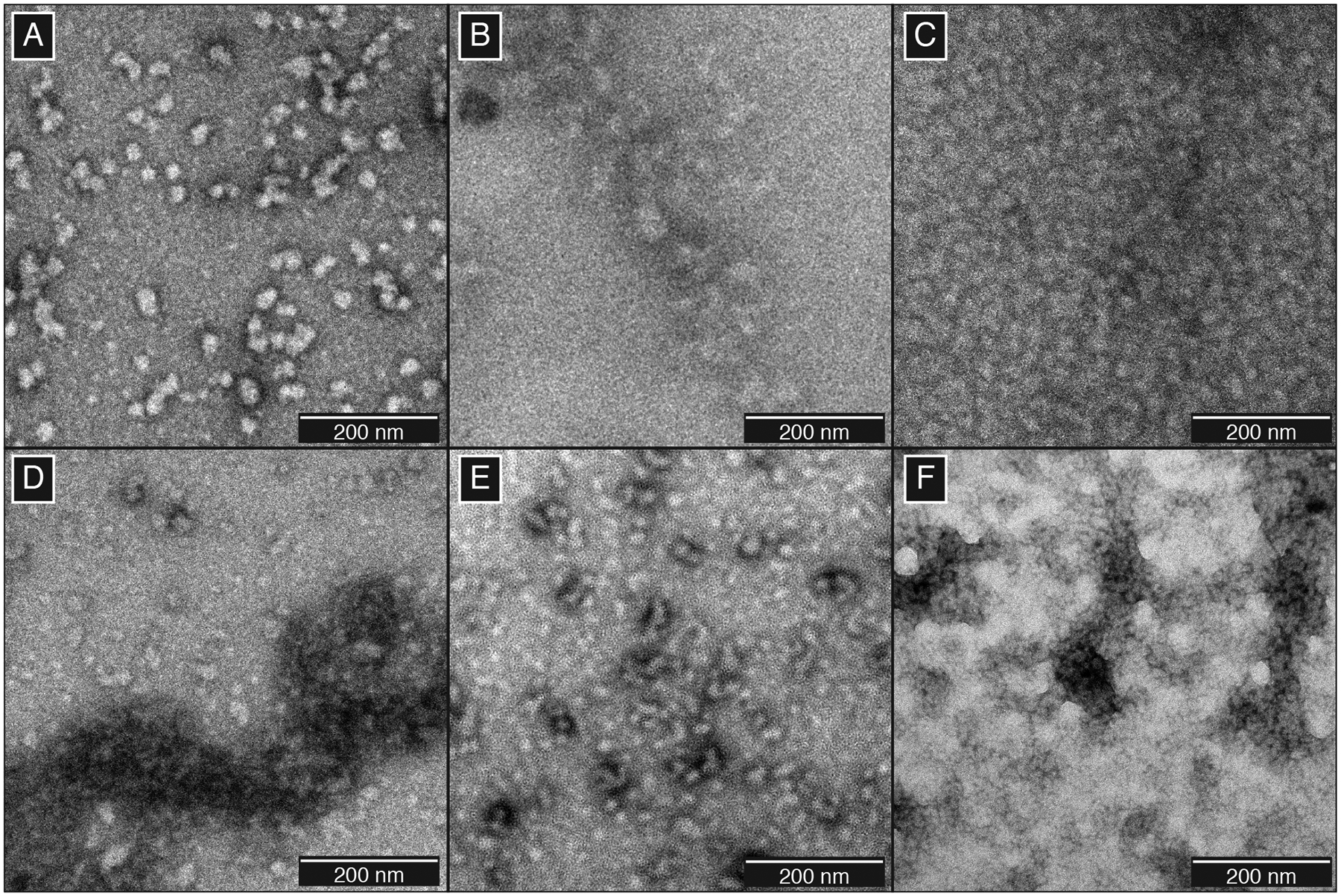
Figure 1:

TEM micrographs of γS-crystallin aggregates. (A) Native aggregates of γS-D26G, (B) γS-G18V, and (C) γS-V42M. Photodamaged aggregates formed using (D) UVA or (E) UVB radiation. (F) Aggregates resulting from the treatment of 0.5 equivalents of CuCl2.
