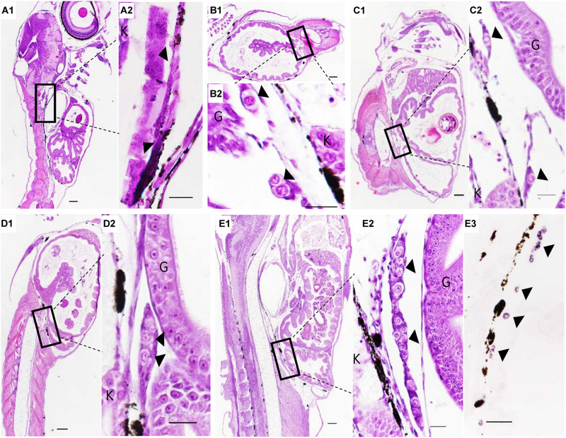
FIGURE 3.
The PGCs migration after birth of Sebastes schlegelii. (A1–E2) were tissue sections with H&E stain. (E3) was SISH corresponding to (E2). PGCs were located in the upper part of the body cavity under the notochord at 2 dab, sagittal sections (A1,A2) and covered by a single-layer somatic cell at 8 dab, coronal sections (B1,B2). PGCs migrated to the genital ridge at 9 dab, sagittal sections (C1,C2) and formed elongated gonadal primordia at 10 dab, sagittal sections (D1,D2). Distinct primordial gonads were discovered in the posterior part of the body cavity (E1,E2), and the PGCs were strongly stained with the Ssvas antisense probe by SISH at 15 dab, sagittal sections (E3). Black arrowheads indicated PGCs; G and K indicated gut and kidney, respectively. Scale bar, 100 μm (A1,B1,C1,D1,E1); 50 μm (A2,B2,C2,D2,E2,E3).

