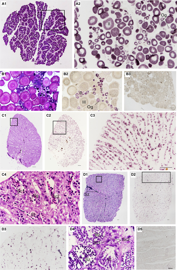
FIGURE 5.
Distribution of Ssvas transcripts in gonad germ cells by SISH. Paraffin section and H&E of ovaries in 1-year-old (A1) and sexually mature black rockfish (B1), respectively. (A2) Adjacent tissue section of the black box in (A1) was hybridized with the Ssvas antisense probe. (B2) Adjacent tissue section of (B1) was hybridized with the Ssvas antisense probe. (B3) The paraffin section of the ovary was hybridized with the Ssvas sense probe. The paraffin section and H&E of testis in 1-year-old (C1) and sexually mature black rockfish (D1), respectively. (C2,D2) Adjacent tissue sections of (C1,D1) were hybridized with the Ssvas antisense probe, respectively. (C3,D3) The high-magnification image of the black boxes in (C2,D2), respectively. (C4–D4) The high-magnification image of the black boxes in (C1,D1), respectively. (D5) The paraffin section of testis was hybridized with the Ssvas sense probe. Og, oogonia; I, early previtellogenic phase; II, late previtellogenic phase; III, early vitellogenic phase; Sg, spermatogonia; Sc1, primary spermatocytes; Sc2, secondary spermatocytes; St, spermatids; Sz, spermatozoon. Scale bar, 100 μm (A1–C4,D3–D5); 200 μm (D1,D2).

