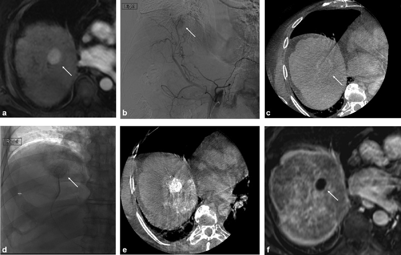
Fig. 1.

Imaging of conventional transarterial chemoembolization treatment with intraprocedural cone-beam computed tomography (CBCT) guidance. Pretreatment magnetic resonance imaging (MRI) reveals an arterially hypervascular tumor in segment 8 of the liver (white arrow) ( a ). Angiographyrevealed tumor blush in the expected location (white arrow) ( b ). Intra procedural CBCT was performed, revealing washout on the delayed imaging phase (white arrow) ( c ). The feeding vessel to the tumor was selected and embolized using doxorubicin emulsified with Lipiodol followed by polyvinyl alcohol particles. Preferential Lipiodol uptake by the tumor can be noted on both angiography (white arrow) ( d ) and CBCT ( e ). Follow-up MRI 1 month after the procedure demonstrates complete response with absence of any residual contrast enhancement (white arrow) ( f ).
