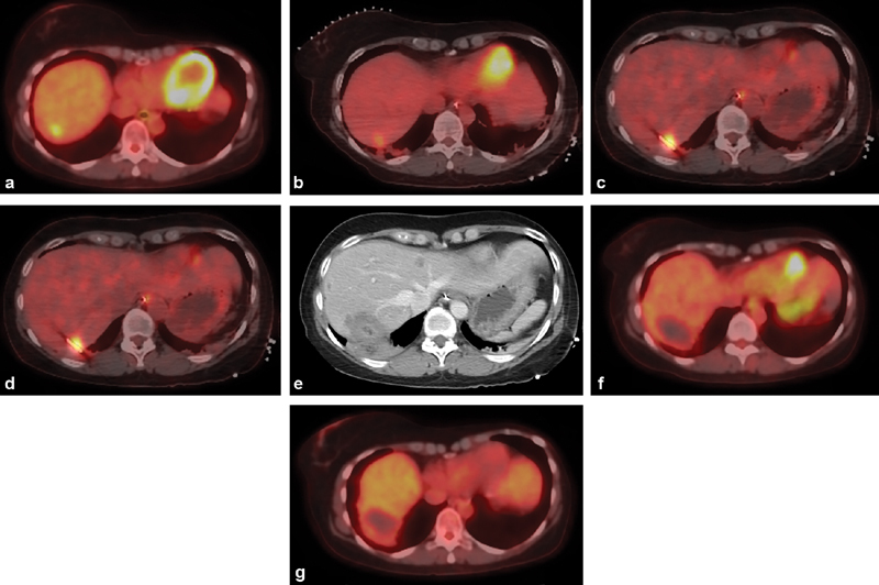
Fig. 2.

Percutaneous PET/CT-guided microwave ablation. ( a ) Axial image from a PET/CT in a 48-year-old woman with locally advanced hormone receptor-positive breast cancer shows a solitary FDG-avid liver metastasis in the hepatic dome, which developed 10 years after tamoxifen therapy. ( b ) Intraprocedure preablation PET/CT is obtained after administration of 4 mCi of FDG. ( c ) Once the ablation probe is placed, a repeat PET/CT is obtained to demonstrate positioning of the probe within the tumor. ( d ) Once ablation is complete, an additional 8-mCi FDG is administered and a final intraprocedure postablation PET/CT is obtained to demonstrate eradication of FDG-avid tumor. This imaging is performed 30 minutes after FDG administration; during that period, a contrast-enhanced CT is obtained to demonstrate the ablation zone ( e ). Follow-up PET/CT performed 6 weeks ( f ) and 6 months ( g ) later demonstrate no residual, recurrent, or new hepatic metastasis.
