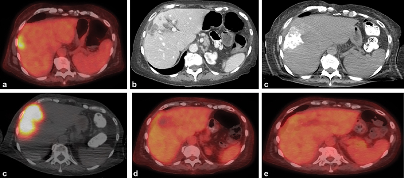
Fig. 3.

Radiation segmentectomy for solitary liver metastasis. ( a ) Axial image from PET/CT in a 69-year-old woman with hormone receptor-positive invasive breast cancer shows a solitary liver metastasis. ( b ) Axial image from a contrast-enhanced CT 2 months later demonstrates that tumor grew over 2 months despite systemic therapy. Liver biopsy demonstrated that the liver metastasis was HER2-negative, in contrast to the primary cancer. ( c ) Intraprocedural CTA identified the segmental arteries supplying the tumor, allowing for a segmental treatment. ( d ) Postadministration SPECT/CT demonstrates dense distribution of yttrium-90 within the tumor. Axial PET/CT images at 1 month ( e ) and 4 months ( f ) demonstrate complete response with no residual disease.
