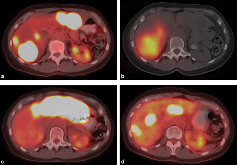
Fig. 4.

Radioembolization of multifocal liver metastasis. (a) Axial image from PET/CT in a 57-year-old woman with hormone receptor-positive breast cancer shows multifocal bilobar liver metastasis progressing despite several lines of systemic therapy. ( b ) Axial SPECT/CT image obtained immediately following right lobar radioembolization demonstrates distribution of yttrium-90 within right hepatic metastases. ( c ) Axial image from PET/CT 2 months later demonstrates complete response in right hepatic metastases, but progression in left lobar metastases, with subsequent left lobar radioembolization. ( d ) Axial image from PET/CT 2 months after left lobar radioembolization demonstrates partial response in left hepatic metastases, with interval regrowth in the right lobe.
