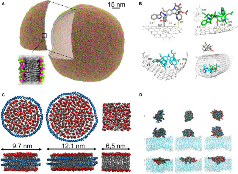
FIGURE 5.

Nanoparticles. (A) Liposome simulated with dry MARTINI model, reproduced with permission from Arnarez et al. (2015), Copyright (2015) American Chemical Society; (B) carbon nanotube used for delivery of vinblastine (Li et al., 2016a), Copyright (2016) American Chemical Society; (C) nanodiscs formed of POPC and membrane scaffold protein MSP1D1 (Left), MSP1E3D1 (middle), and lipid bilayer (right), protein is shown as blue ribbon, phosphate groups of lipids shown as red sphere, and acyl tail as gray sticks, reproduced with permission from Stepien et al. (2020); (D) PAMAM dendrimer in water phase (top), at the lipid bilayer in gel phase (middle), and at the lipid bilayer in fluid phase (down), reproduced with permission from Kelly et al. (2008), Copyright (2008) American Chemical Society.
