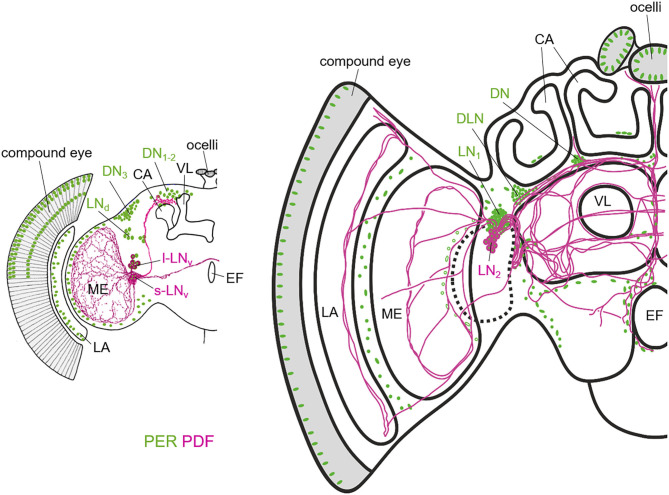
Figure 2.
Schematic representation of the PER and PDF expressing neurons in the fruit fly (left) and honey bee brain (right). The two brains differ in overall size, but especially in the size of the mushroom bodies of which the Calyces (CA) and the vertical lobes (VL) are highlighted. The vertical lobe is also called α-lobe in Drosophila. While the honey bee has two large calyces per brain hemisphere, the fruit fly has only one rather small one. In Drosophila, the only PDF-fibers that run into the dorsal brain, terminate anterior, and slightly dorsally of the CA. In the honey bee, the PDF fibers terminate anterior and ventrally of the CAs and they run into many more central brain areas than in Drosophila. PER is not only present in the lateral (LN) and dorsal neurons, but also in the photoreceptor cells of the compound eyes and ocelli and in numerous glia cells that are only indicated in the lamina (LA) and the distal medulla (ME) in Drosophila and in few other brain regions in the honey bee. Note that the nuclei of the photoreceptor cells are only shown in the dorsal half of the compound eye in Drosophila. The honey bee scheme is modified from Beer et al. (2018). For details see text.

