
Figure 4.
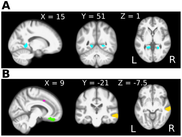
Gray matter volume differences associated with hearing loss and with tinnitus. Threshold free cluster enhancement (TFCE) was employed to investigate group differences in modulated gray matter images. (A) Comparison between the hearing loss groups, with and without tinnitus. In the group with tinnitus, larger gray matter volume of the bilateral lingual gyri was observed. These areas are depicted in blue on a medial view of the sagittal plane of the right hemisphere, and a coronal and axial cross section of the entire brain. (B) Comparison between the hearing loss group without tinnitus and the control group. In the hearing loss group without tinnitus less gray matter volume was observed in the cingulate cortex (pink), the orbitofrontal cortex (green), and the middle temporal gyri (yellow), depicted on a medial view of the sagittal plane of the right hemisphere. Additional clusters with less gray matter volume, not shown here, were observed in the superior and inferior temporal gyri and the fusiform gyrus (see Table 4). The comparison between the hearing loss group with tinnitus and the controls revealed no significant differences and is therefore not shown in this figure.