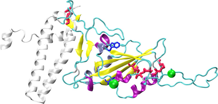
Figure 1.
Structure of SARS-CoV-2 Receptor Binding Domain bound to ACE2. The secondary structure elements of RBD are differentially colored (Alpha helices: purple, 3_10 helices: iceblue, beta strands: yellow, and turns/coil: cyan). Disulfide bridges (red) and tryptophan residues (blue) are shown as sticks, while N-glycosylation asparagine residues (green) are shown as VDW spheres. The region of ACE2 encompassing residues 1–115 (colored white) which interacts with RBD is also shown. The structure was generated using Protein Data Bank (PDB) structures 6xm0 and 6m0j.

