Figure 7.

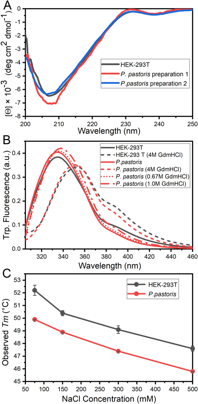
Conformation and stability of different purified RBD forms characterized by circular dichroism (CD) Spectroscopy, Tryptophan Fluorescence and Temperature-induced Denaturation. (A) Far-UV CD spectra of RBD produced in HEK-293T cells (black), and two different preparations of RBD produced in P. pastoris (red and blue). (B) Tryptophan fluorescence emission was monitored by excitation at 295 nm in 20 mM Tris–HCl, 100 mM NaCl, pH 7.0 at 25 °C. The spectra of RBD obtained in HEK-293 T (black) and in P. pastoris (red) are shown in native conditions (solid line) and in the presence of 4.0 M GdmCl (dashed line) after a 3 h incubation. Refolding of RBD produced in P. pastoris was performed by dilution to final concentrations of 0.7 M (red dot line) and 1.0 M (red dash-dot line) GdmCl. (C) Stability analysis of RBD. Temperature-induced denaturation of RBD produced in P. pastoris (red) and HEK-293T cells (black) under different ionic strength conditions (75, 150, 300 and 500 mM NaCl) was followed by Sypro-orange fluorescence.
