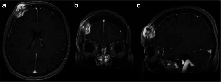
Fig. 1.
a Axial, b coronal, and c sagittal T1-weighted post-contrast MRI with right frontal bone mass measuring 3.2 × 2.2 cm in diameter with extension to the subcutaneous scalp, as well as intracranial extension into the epidural space. No evidence of brain parenchymal involvement or vasogenic edema was noted

