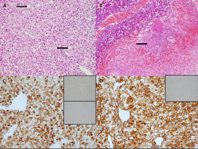
Figure 1.
Histology from resection of liver lesion in first case. Large polygonal tumour cells with abundant cytoplasm were arranged around ectatic vessels. occasional fat-containing cells were present (arrow) (A, H&E ×200 magnification). Fewer than 1 mitosis was identified per 10 high power fields. Foci of tumour necrosis were present (B, arrow, ×100 magnification). The tumour expressed HMB-45 (C, main image, HMB-45 immunostain ×200 magnification) and also expressed smooth muscle actin (D, main image, SMA immunostain ×200 magnification). There was no expression of S100 (C, upper inset, S100 immunostain ×200 magnification), no expression of OCH1E5 (C, lower inset, OCH1E5 immunostain ×100 magnification) and no expression of desmin (D, inset, desmin immunostain ×100 magnification).

